Gene Editing
Gene Editing
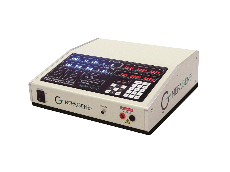
Super Electroporator NEPA21 Type II
- Novel 4-Step Multiple Electroporation Pulse.
- It is especially suitable for the difficult-to-transfect cells, ex vivo and in vivo animal tissues.
- Suitable for gene editing of embryo.
- High transfection efficiency and high cell viability.
- The parameters of the electroporation pulse are visible, adjustable and have wide applicability.
- Without special buffers and low operational costs.
Super Electroporator NEPA21 Type II is suitable for in vitro and in vivo transfection.
Make transfection easier and Free.
Super Electroporator NEPA21 Type II adopts a newly designed electroporation pulse, which is specially designed for the difficult-to-transfect cells, microalgae, fertilized eggs, ex vivo and in vivo animal tissues, and has the advantages of high transfection efficiency and high cell viability.
Product Details:
In 2011, NEPAGENE launched a new generation of versatile and highly effective gene transfection systems---Super Electroporator NEPA21 Type II. Compared with the traditional electroporators, NEPA21 adopts the novel 4-step multiple electroporation pulse, which is especially suitable for the difficult-to-transfect cells, ex vivo and in vivo animal tissues. With the patented voltage decay design, NEPA21 can not only achieve high transfection efficiency, but also improve cell viability.
Key Features of NEPA21 Electroporation System:
- Novel 4-Step Multiple Electroporation Pulse.
- It is especially suitable for the difficult-to-transfect cells, ex vivo and in vivo animal tissues.
- Suitable for gene editing of embryo.
- High transfection efficiency and high cell viability.
- The parameters of the electroporation pulse are visible, adjustable and have wide applicability.
- Without special buffers and low operational costs.
Application:
- Suspension transfection
Scope of application: primary cells, stem cells, and various difficult transfection cells (such as immune cells, blood cells, etc.).
- Adhesion transfection
Scope of application: adherent cells can be directly transfected, eliminating the steps of cell digestion and re- adherent (Which is very important for improving the viability of some kinds of cells)!
- In vitro
Scope of application: tissue section, brain section, organ, embryo, etc
- In vivo
Scope of application: brain, retina, muscle, skin, liver, kidney, testicles, etc
The production of NEPA21 series electroporation instrument, ECFG21 high-efficiency cell electrofusion and electroporation instrument and PicoPipet single-cell separation and extraction system has been well-known in the research field and has been cited by hundreds of literatures, including high-level journal articles, such as Nature, Cell, PNAS, Genes & Dvelopment, etc.
<!--[if !supportLists]-->Ø <!--[endif]-->Hendriks D, Pagliaro A,
Andreatta F, et al. Human fetal brain self-organizes into long-term expanding
organoids[J]. Cell, 2024.
<!--[if !supportLists]-->Ø <!--[endif]-->Diaz-Cuadros M, Wagner D E,
Budjan C, et al. In vitro characterization of the human segmentation clock[J]. Nature, 2020, 580(7801): 113-118.
<!--[if !supportLists]-->Ø <!--[endif]-->Nakazawa Y, Hara Y, Oka Y, et
al. Ubiquitination of DNA damage-stalled RNAPII promotes transcription-coupled
repair[J]. Cell, 2020, 180(6):
1228-1244. e24.
<!--[if !supportLists]-->Ø <!--[endif]-->Barnabe-Heider et al. Genetic
manipulation of adult mouse neurogenic niches by in vivo electroporation. Nature Methods, 2008 Feb;5(2):189-96.
<!--[if !supportLists]-->Ø <!--[endif]-->Limura T and Pourquie O.
Collinear activation of Hoxb genes during gastrulation is linked to mesoderm
cell ingression. Nature, 2006 Aug
3;442(7102):568-71.
<!--[if !supportLists]-->Ø <!--[endif]-->Sanada K and Tsai LH. G
Protein betagamma Subunits and AGS3 Control Spindle Orientation and Asymmetric
Cell Fate of Cerebral Cortical Progenitors. Cell, 2005 Jul 15;122(1):119-31
<!--[if !supportLists]-->Ø <!--[endif]-->Ladher RK et al. FGF8
initiates inner ear induction in chick and mouse. Genes and Development, 2005 Mar 1;19(5):603-13.


